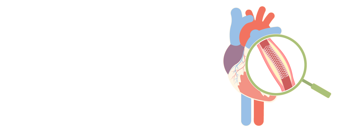
Heart blockage, also known as coronary artery disease, or CAD, is an important health issue worldwide. For quick action and the avoidance of effects, early identification of CAD is essential. Non-invasive techniques are essential for diagnosing CAD in patients, even if angiography is still the standard of care. Without the discomfort of traditional angiography, these techniques—echocardiography, stress testing, cardiac MRI, CT angiography, and myocardial perfusion imaging—offer valuable details about the body, blood flow, and function of the heart. The basis and importance of non-invasive methods for detecting heart blockages are looked at in this blog.
Electrocardiogram (ECG)
- ECG is a fundamental tool for assessing cardiac rhythm and detecting abnormalities indicative of heart blockage.
- It records the electrical activity of the heart, providing information about heart rate, rhythm disturbances, and ischemic changes.
- Specific ECG patterns such as ST-segment depression or elevation, T-wave inversion, and bundle branch blocks may suggest underlying CAD.
- While ECG alone may not definitively diagnose CAD, it serves as a valuable initial screening tool.
Echocardiography
- Echocardiography utilizes sound waves to create images of the heart, allowing visualization of its structures and function.
- Doppler echocardiography assesses blood flow within the heart chambers and can detect abnormalities such as wall motion abnormalities or valve dysfunction associated with CAD.
- Stress echocardiography combines echocardiography with physical or pharmacological stress to evaluate cardiac function under conditions of increased demand, aiding in the detection of ischemia and CAD.
Stress Testing
- Stress testing involves inducing physical or pharmacological stress while monitoring the heart's response through ECG, echocardiography, or nuclear imaging.
- Exercise stress testing, typically performed on a treadmill or stationary bike, assesses the heart's response to increased workload.
- Pharmacological stress testing involves administering medications that simulate exercise-induced stress when physical activity is contraindicated.
- Stress testing helps evaluate symptoms suggestive of CAD, assess exercise capacity, and identify ischemic changes indicative of heart blockage.
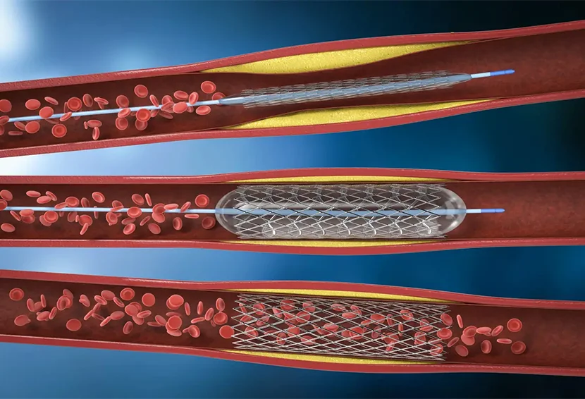
CT Angiography (CTA)
- CTA utilizes computed tomography (CT) imaging to visualize the coronary arteries and assess for the presence of blockages or stenosis.
- It provides detailed anatomical information, allowing for the identification of plaque buildup and assessment of the extent and severity of CAD.
- CTA is less invasive than traditional angiography and can effectively rule out significant coronary artery disease in many cases.
- However, it exposes patients to radiation and may not be suitable for individuals with kidney impairment or allergies to contrast dye.
Magnetic Resonance Imaging (MRI)
- Cardiac MRI offers a comprehensive assessment of cardiac structure, function, and perfusion without exposure to ionizing radiation.
- It can detect myocardial ischemia, assess myocardial viability, and evaluate for the presence of scar tissue secondary to previous myocardial infarction.
- MRI with contrast enhancement provides dynamic imaging of myocardial perfusion, aiding in the detection of CAD-related abnormalities.
Myocardial Perfusion Imaging
- Myocardial perfusion imaging (MPI) involves injecting a radioactive tracer and imaging its distribution within the myocardium at rest and during stress.
- Techniques include single-photon emission computed tomography (SPECT) and positron emission tomography (PET).
- MPI helps identify areas of reduced blood flow indicative of ischemia or infarction, guiding the diagnosis and management of CAD.
Conclusion
In conclusion, non-invasive techniques are essential for recognizing and evaluating heart blockages because they provide important diagnostic data without the risks of invasive tests like angiography. The complete approach to detecting CAD which includes electrocardiography, echocardiography, stress testing, CT angiography, cardiac MRI, & myocardial perfusion imaging enables immediate action and better outcomes for patients. Although angiography is still necessary in some situations, non-invasive methods are developing and becoming more accessible and safe for the diagnosis and treatment of heart blockages.
Written and Verified by:
Dr. Gurkirat Singh Sidhu
Years of Experience : 10 Years Of Experience
Senior Consultant Cardiology Department
Meet the doctor


_.webp)