An angiography includes injecting contrast dye into your body so your doctor can see it with an X-ray machine. Images on a screen show blood flow and blockages in your blood vessels.
Before your angiography test, your doctor may want to check your blood to see how well it clots. They also want to make sure your kidneys are performing properly.
Follow these instructions after midnight the night before your exam:
If your healthcare professional approves:
Follow all the instructions given by your healthcare provider on the day of your angiography:
During the test, the medical team ensures you are comfortable and monitors your vital signs closely as the angiography procedure begins:
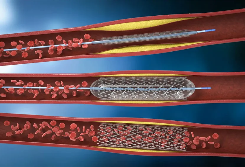
If your doctor discovers a blockage, he or she may perform an angioplasty immediately. This method uses a small balloon to press the blockage against the arterial wall. An angioplasty may be all you need if it improves blood flow and leaves less than 30% of the blockage after the treatment.
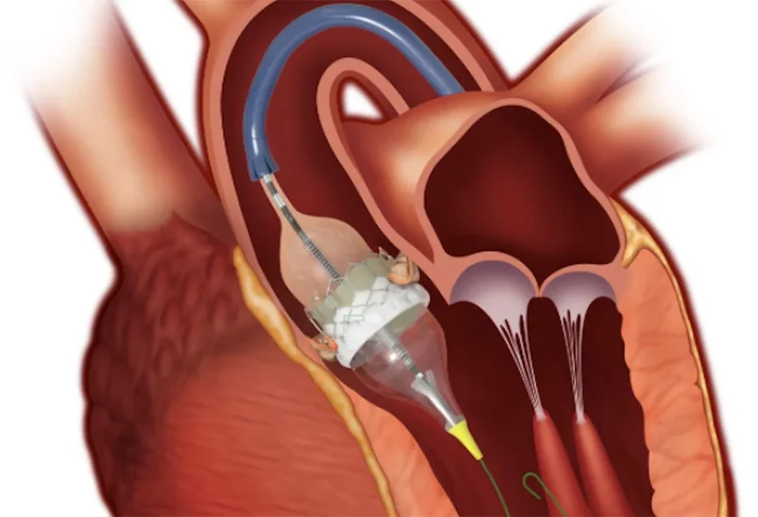
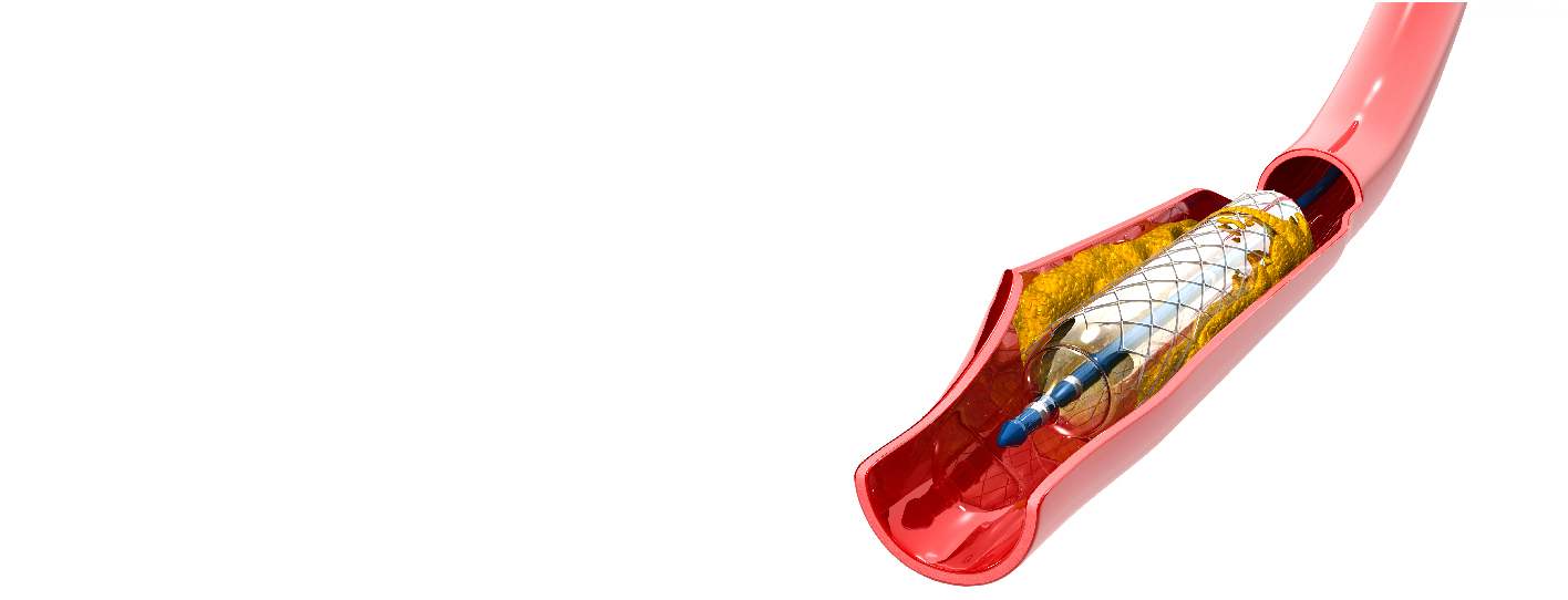
If an angioplasty fails to produce a large enough opening for blood to pass through, you may require a stent. This small metal tube remains inside your blood vessel to keep it open. Your provider can perform this immediately following your angioplasty.
If a stent is also not best suited for an individual then another technique, bypass surgery is recommended in that case.
Your healthcare practitioner will remove the catheter and bandage the punctured area in your skin. They will apply pressure to the bandaged area for at least 15 minutes to stop or prevent bleeding. If doctors insert the catheter through your leg, you will need to be in bed for four to six hours. This reduces the likelihood that your incision will bleed. Before you leave for home, your provider will assess you and go through at-home instructions.
Also, read: How to Check Heart Blockage Without Angiography?
You should be able to leave the hospital on the same day as your angiography. After you return home, don't lift anything more than 4 kg or stoop or bend for the following two days. This should prevent the incision from bleeding. Some patients may need to spend the night in the hospital following their angiography procedure.
The potential risks associated with angiography are low. However, complications can occur in the area where your provider punctured your skin to reach your artery. Angiography problems occur in less than one percent of cases. The risks of an angiography procedure usually involve your puncture site and consist of:
Complications of angiography, though rare, can arise and may include reactions to contrast dye or issues related to the catheter procedure. Here's an overview:
Minor adverse events include a small amount of bleeding or bruising at the access site or a small hematoma. A hematoma is a contained collection of clotted blood outside the artery. Access site hematomas are usually small and require no specific therapy. They occur in less than 3% of patients.
A hematoma is considered a major complication if any of the following occurs: a blood transfusion is required; surgical evacuation is necessary; readmission to the hospital; prolonged hospital stay. Major bleeding complications are rare and have not yet been reported in UFE patients.
Artery walls can suffer injury during entry and further administration of the catheter (small plastic tube). Serious problems that require hospitalization, surgery, other invasive interventions, or hospital readmission are rare.
Iodinated radiography contrast can cause temporary or chronic renal failure. This usually occurs in patients who already have problems with kidney function, such as diabetics. The tiny amount of contrast utilized during the test does not seem to pose a risk of renal failure in otherwise healthy people.
Some people are allergic to contrast agents. Usually, this is mild, with the most common symptoms being a rash or hives that resolve quickly in most cases. Approximately 1 in 4000 people will experience a more significant reaction, such as severe asthma or airway swelling, requiring intravenous treatment and an extended period of care. An extreme (anaphylactic) reaction that ends in life loss is extremely rare.
Your healthcare professional may discover minor blockages that do not require additional treatment. You may need to take medicine or make adjustments to your usual routine. For example, you may need to increase your physical activity or eat healthier. You may have larger blockages requiring more extensive therapy. Your doctor may perform an angioplasty and insert a stent to keep your artery open. They can perform this during your angiography procedure.
Some patients require an additional procedure on another day. Your doctor may propose a bypass procedure that allows blood to flow around the blocked area.
Also, read: How to Remove Heart Blockage without Surgery?
Angiography is one of the most effective tools for diagnosing and treating vascular disorders. It provides a detailed view of blood vessels and enables healthcare professionals to detect blockage, any abnormalities, and blood flow problems. Early detection and quick intervention have led to saving lives and improved outcomes.
Q1: Is angiography painful?
A: A long, thin, flexible tube (catheter) is placed into the artery and gently directed to the area under examination; you may feel some pushing and pulling during this process, but it should not be painful.
Q2: Can angiography clear a blockage?
A: A method called angioplasty may be used to treat narrowed coronary arteries during an angiography. To remove the blockage, a special catheter is inserted through the blood channels and into the coronary arteries. Another surgical option for severely narrowed coronary arteries is bypass surgery.
Q3: When should I call my doctor?
A: Please contact your healthcare provider if you have:
Q4: Who performs an angiography?
A: An interventional cardiologist performs an angiography. Interventional radiologists and vascular surgeons perform angiography of peripheral arteries. Interventional neurologists perform cerebral angiography. The provider who performs the angiography can study your images and identify problem areas.

Consultant Cardiologist Cardiology Department
Meet the doctorFrom Recent Advancements in Heart Care to Tips and Tricks to make your Heart Healthy Again, stay updated with reliable and informative blogs by our experts.


 Causes, Symptoms, Diagnosis & Treatment_.webp)