

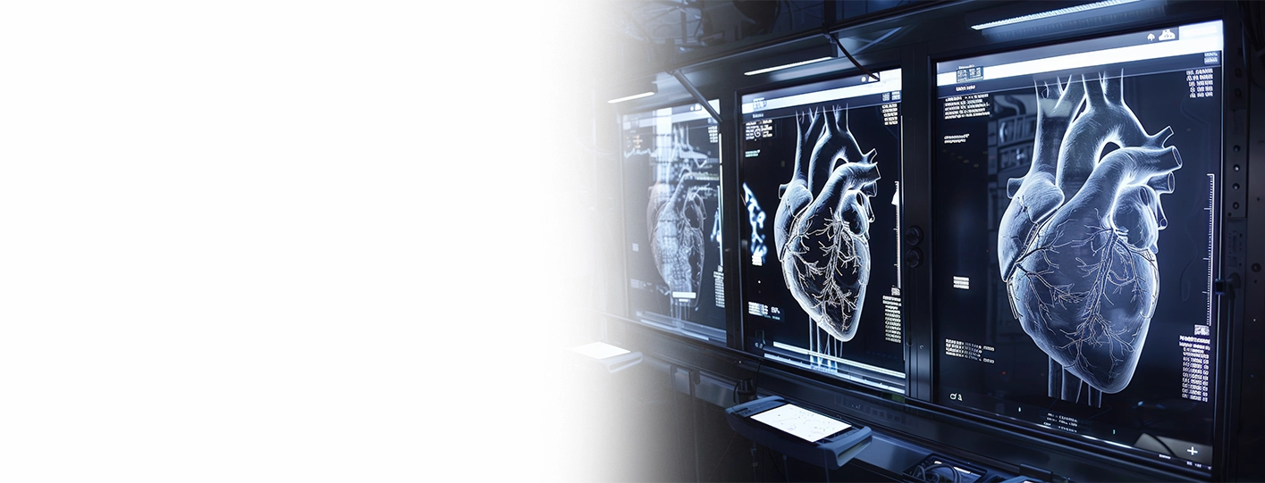
An echocardiogram is an imaging technique that uses sound waves to take moving pictures of your heart. It works very similarly to an ultrasound scan done during pregnancy, which helps to record the movement of the fetus. The process does not require any injectables, and no radiation is involved. It is simple, hassle-free and generally painless.
The procedure is quite helpful in diagnosing and assessing conditions like heart failure, heart disease and congenital heart diseases.
It can be of several types. The most common of which is a transthoracic echocardiogram, or simply TTE. Other types include stress echo and transoesophageal echo (TOE).
3D echocardiogram is like an upgraded form of the standard echocardiogram. What makes it special is the use of a matrix-array probe that can scan the whole heart in one go. This generates sound waves in a pyramid-shaped pattern that helps to create a three-dimensional image of the whole heart in real time.
Let's try to understand this in a simple way. Imagine trying to understand a whole building by looking at the photograph of just one floor. You can see a wall, a window, maybe some decorative items lying nearby, but you do not know what the room actually looks like. Your vision can't go beyond that photograph.
This is how a standard echocardiogram works. It gives detailed images of a part of the heart; however, when you want to assess the whole heart, and not just a part of it, you would need a more detailed view. This is exactly what 3D echocardiography does. It allows doctors to look at the heart from different angles. They can rotate it, slice it, and measure it from any direction.
| Feature | Standard 2D echocardiogram | 3D echocardiography |
| Image | It gives a flat, slice-like view that can only be seen from the front | It gives a three-dimensional view that can be seen from any angle |
| Valve shape | Doctors assume the 3-D shape, but cannot actually see it | Doctors can see the valve shape in 3D |
| Heart chamber size | This is estimated using geometric assumptions | This is precisely measured, directly from the volume |
| Radiation | None | None |
| Availability | Widely available | Needs special equipment and training |
| Best used for | Routine screening and basic function | Complex valve conditions, surgical planning, precise measurements |
In case of 3D transoesophageal echo, you may be required to fast for a couple of hours before the test. This is because the probe is passed through the food pipe. The throat is numbed using mild sedation.
The pumping strength of the heart is usually reported as ejection fraction or EF. The 2019 review published in the Heart, Lung and Circulation states that “3D echo has consistently shown superior accuracy and reproducibility than 2D echo in the assessment of left ventricular (LV) volumes and ejection fraction”
The mitral valve has a complex shape with non-flat leaflets; however, 3D echo can easily help to assess its structures as well as the surrounding tissues, according to a review in Mitral Valve Anatomy And Function: New Insights From Three-Dimensional Echocardiography
In patients suffering from heart valve disease, this can help to locate the leaking part of the valve, measure valve opening in mitral valve stenosis, see whether valve flaps are fused and plan the proper course of surgery.
A review report on 3D Echo in Routine Clinical Practice Published in 2019 states that 3D echo is superior to 2D echo for atrial septal defects. This is because it helps to give a face-on view that helps to assess the shape, size, and number of defects accurately.
A review published in the Journal of the Indian Academy of Echocardiography & Cardiovascular Imaging highlights that 3D echocardiography has helped interventional cardiologists to perform complex procedures like balloon mitral valvuloplasty and paravalvular leak closure by allowing precise assessment before, during and after the procedures.
By now you may have understood how 3D Echo helps in the assessment and treatment of heart valve diseases. However, this can also be used for assessing other conditions like:
3D echocardiography may also be used before or after a surgical procedure to plan the treatment and how the patients are responding to it. It may also be used for long-term monitoring of patients, especially those who are undergoing chemotherapy or those with leaky valves.
Just like other medical procedures, 3D echocardiography has certain limitations as well.
You should immediately see a cardiologist if you're experiencing symptoms like breathlessness, chest discomfort, palpitations, dizziness, fainting, swollen feet, and a new murmur. People who have a history of rheumatic fever should go for regular checkups. Emergency medical care is needed in case of chest pain, sudden breathlessness, and fainting.
3D echocardiography does offer some benefits over the standard echocardiogram by giving a more detailed view. It can sharpen the diagnosis of valve diseases as well as help in planning the treatment. The procedure is offered at Artemis Hospital. You can book an appointment today and get yourself evaluated by our experts.
Q1. What is 3D echocardiography?
A: 3D Echocardiography is a technique that helps to create moving 3D images of the heart from various angles for precise diagnosis and assessment.
Q2. How is 3D echo different from a regular echocardiogram?
A: A 3D echo is different from a regular echocardiogram as the latter helps to capture only 2D slices, whereas the former can capture the volume of the heart and measure it directly.
Q3. Is 3D echocardiography painful?
A: A chest-based 3D echocardiography is not painful; however, you may feel some discomfort while going for a transoesophageal 3D scan since the probe is introduced through the throat.
Q4. Does 3D echo use radiation?
A: No, 3D echo does not involve the use of radiation. It only uses sound waves.
Q5. When is 3D echocardiography used in heart valve disease?
A: 3D echocardiography is used in heart \valve diseases when the doctors want to assess the valve shape, measure mitral valve area, find the source of a leak and plan repair or replacement.
This article is for educational purposes only. It is not medical advice and does not replace diagnosis or treatment by a qualified cardiologist. Imaging choices depend on individual assessment, and no outcome can be guaranteed. Please consult a registered medical practitioner about any heart symptoms, and seek emergency care for severe chest pain or breathlessness.
From Recent Advancements in Heart Care to Tips and Tricks to make your Heart Healthy Again, stay updated with reliable and informative blogs by our experts.

 WhatsApp Us (24/7)
 +91 9070902010 (24/7)


 Previous



.webp)
